
Perivascular epithelioid cell tumors, or PEComas, represent a family of mesenchymal tumors characterized by the simultaneous expression of melanocytic and muscular markers.
These tumors can arise in various anatomical sites, including the kidney, lung, bladder, prostate, pancreas, liver, falciform ligament/round ligament of the liver, breast, skin, eyes, skull base, colon, soft tissues, retroperitoneum, and the female genital tract.
Gynecological PEComas are rare, constituting approximately 25 to 30% of cases of this tumor type, with the uterine body accounting for the majority of cases (close to 72% in the retrospective study by Liu et al. (2019).
This study aims to report a case of malignant PEComa of the uterine cervix, focusing on the rarity of this tumor type, the proposed surgical treatment, the pathologist’s importance in the differential diagnosis of this neoplasm, and the follow up of this patient.
A 34-year-old female patient was referred by a gynecologist to a surgical oncology service due to vaginal bleeding and an expansive mass in the uterine cervix evolving over 5 months.
The patient had 2 previous gestations. She was using oral contraceptives and had a history of discontinuing them due to vaginal bleeding. A colpocitology had been previously conducted (1 month prior to the consultation with surgical oncology team), revealing cervical intraepithelial neoplasia (CIN) II/III. Upon speculum examination, a large friable, bleeding, exophytic mass with necrotic areas in the uterine cervix was observed, occupying the entire lateral aspect of the vaginal canal, rendering the cervical orifice unidentifiable.
Biopsy samples were collected from the mass, and the histopathological report indicated a poorly differentiated carcinoma, featuring clear and signetring-like cells, requiring immunohistochemistry (IHC) to confirm the lesion’s origin.
IHC was positive for estrogen receptor (EP1), desmin (D33) in rare cells, focal alpha-smooth muscle actin (1A4), weak and focal melan-A (A 103), HMB45, TFE3 (EP285 clone), weak p63 (DAK-p63 clone) in rare cells, vimentin (V9), and Ki-67 (MIB-1) in approximately 20% of cells, consisting with PEComa.
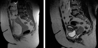
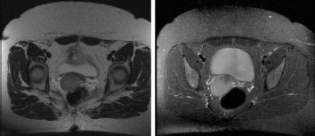
Pelvic magnetic resonance imaging (MRI) revealed a solid ovoid lesion measuring 5 cm in diameter, involving the uterine cervix and extending approximately 3 cm into the vaginal canal and no signs of parametria, rectum or bladder infiltration. (
Figure 1. Sagittal MRI section of the pelvis showing an expansive lesion in the uterine cervix, ovoid, in continuity with the vaginal vault, without evidence of involvement of the parametrium and regional lymph nodes.
Figure 2. Axial MRI section of the pelvis showing an expansive lesion in the cervix, with a cleavage plane with the bladder and rectum.
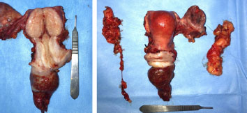
The patient was submitted to surgical treatment. The initial idea was preserve both ovaries due to patient age and histology, but in the transoperative it was observed that the right ovary had an increase in volume and a suspicious appearance for neoplastic infiltration, despite the normal appearance at MRI. We performed a total hysterectomy with unilateral salpingo-oophorectomy and bilateral pelvic lymphadenectomy (
Figure 3. Product of total hysterectomy with unilateral salpin- go-oophorectomy and bilateral pelvic lymphadenectomy.
Postoperatively, the patient showed good progress and was referred for consultations with clinical oncology and radiation oncology to discuss adjuvant therapy options. It was decided not to proceed with any form of adjuvant therapy for this patient, who continues under clinical follow up, showing no evidence of disease after 28 months.
Gynecological PEComas are rare, with the majority being sporadic tumors, and a smaller percentage associated with complex tuberous sclerosis (CTS). PEComas of the uterine cervix are even rarer - only 16 cases have been reported in the literature until now.
| Cases | Age | History of CTS/gene mutation | Tumor size | Folpe’s criteria for agressive behavior | Treatment | Follow up |
|---|---|---|---|---|---|---|
| Case 1 | 41 | Yes | 2.2 cm | None | Total histerectomy with bilateral salpingo- oophorectomy | 35 months, without evidence of recurrence |
| Fadare et al., (2004) | Intraabdominal PEComatosis | |||||
| Case 2 | 48 | No | 2 cm | High nuclear grade | Local excision and adjuvante radiotherapy | 21 months, without. evidence of recurrence |
| Folpe et al., (2005) | ||||||
| Case 3 | 28 | No | 3 cm | None | Histerectomy and lymphadenectomy | 36 months, without evidence of recurrence |
| Folpe et al., (2005) | ||||||
| Case 4 | 25 | No | Cervical anterior lip ulceration | Infiltrative margins | Total histerectomy with bilateral salpingo- oophorectomy and lymphadenectomy | NI (no information) |
| Azad et al., (2006) | ||||||
| Case 5 | 24 | No | Cervical membranous tissue | Infiltrative margins | Local excision | 2 early cervical recurrences - 4 months after the first procedure, and 7 months after the second procedure |
| Yamamoto et al., (2010) | ||||||
|
|
|
|
|
|
| 12 months after the thirdprocedure: without evidence of recurrence |
| Case 6 | 46 | No | 3-4 cm | None | Total histerectomy with bilateral salpingo- oophorectomy + radiotherapy (external beam radiation therapy - EBRT) + immunotherapy | 36 months, without evidence of recurrence |
| Bradshaw et al., (2010) | ||||||
| Case 7 | 61 | No | 9 cm | Size > 5 cm | mTOR inhibitors | Death (8 months) |
| Wagner et al., (2010) | Pulmonary metastasis at diagnosis | |||||
| Case 8 | 59 | Yes | Non-specified | NI | Total histerectomy with bilateral salpingo- oophorectomy | NI |
| Lim et al., (2011) | ||||||
| Case 9 | 52 | No | 12 cm | Vascular invasion, necrosis, high nuclear grade | Pelvic exenteration with uterus, vagina, bladder and anal canal resection + adjuvante radiotherapy (28 fractions) | 12 months, without evidence of recurrence |
| Natella et al., (2013) | ||||||
| Case 10 | 57 | NI | 3.3 cm | Multiple bizarre giant cells, necrosis, mitotic index 2MF/5QHPF | Total histerectomy with bilateralsalpingo-oophorectomy | NI |
| Zhang et al., (2013) | ||||||
| Case 11 | 41 | Yes | 4 cm Intraabdominal PEComatosis | Infiltrative margins | Total histerectomy with bilateralsalpingo-oophorectomy | 36 months, without evidence of recurrence |
| Çelik et al., (2014) | ||||||
| Case 12 | 34 | No | 9 cm | Size > 5 cm, necrosis, infiltrative margins | Mass resection | Local recurrence after 2 months |
| Liu et al.,(2014) | | Metastatic pelvic lymph nodes after 5months | ||||
| Case 13 | 51 | No | 2.8 cm | None | Total histerectomy with bilateralsalpingo-oophorectomy | SI |
| Tajima and Koda, (2015) | ||||||
| Case 14 | 43 | No | 3 cm | None | Histerectomy | 36 months, without evidence of recurrence |
| Kovac et al., (2018) | | |||||
| Case 15 | 57 | No | 11 cm | Size > 5 cm, infiltrative margins, high nuclear grade, necrosis, mitotic index > 4 MF/10HPF | Total histerectomy with bilateral salpingo-oophorectomy and pelvic lymphadenectomy | 6 months, without evidence of recurrence |
| Mateva et al., (2019) | ||||||
| Case 16 | 55 | SI | 3 cm | None | Total histerectomy with bilateral salpingo-oophorectomy | 24 months, without evidence of recurrence |
| Tang et al.,(2023) | ||||||
| Case 17 | 34 | SI | 5.5 cm | Size > 5 cm | Total histerectomy with unilateralsalpingo-oophorectomy and bilateral pelvic lymphadenectomy | 28 months, without evidence of recurrence |
| Our case |
NI = No information; CTS = Complex tuberous sclerosis.
The first cervical PEComa was described by Fadare et al., in 2004,
While most PEComas are benign, a subgroup demonstrates aggressive behavior.
In 2005, Folpe et al.
| Category | Folpe’s criteria (2005) | Schoolmeester’s criteria (2014) | Folpe’s modified criteria -Conlon et al. (2015) |
|---|---|---|---|
| Benign | None of below criteria: • Size equal or higher 5 cm • Infiltrative growth pattern • High nuclear and cellularity grade • Mitotic index > 1M/50HPF • Necrosis • Vascular invasion | Less than four below criteria: • Size equal or higher 5 cm • High nuclear grade • Mitotic index > 1M/50HPF • Necrosis • Vascular invasion | None or one below criteria: • Infiltrative margins • Size between 5 and 10 cm • Mitotic index 2-3M/50HPF • Lymphatic and vascular invasion |
| Uncertain malignant potential | One of below criteria: • Nuclear pleomorphism/giant multinucleated cells • Size equal or higher 5 cm | One of below criteria: • Isolated important atypia • Size > 10 cm • Mitotic index equal or higher than 4M/50HPF | |
| Malignant | Two or more below criteria: • Size equal or higher 5 cm • Infiltrative growth pattern • High nuclear and cellularity grade • Mitotic index > 1M/50HPF • Necrosis • Vascular invasion | Four or more below criteria: • Size equal or higher 5 cm • High nuclear grade • Mitotic index > 1M/50HPF • Necrosis • Vascular invasion | Any grade of necrosis or two or more above criteria |
Adapted from Mateva et al., (2019)
There is a correlation between the development of PEComas and tuberous sclerosis complex, a rare genetic disease that can lead to the formation of multiple tumors (mostly benign) in various parts of the body, including the brain, skin, kidneys, heart, eyes, and lungs. In tuberous sclerosis, a mutation occurs in the TSC1 and TSC2 genes, located on chromosomes 9 and 16, respectively. In PEComas, mutations in the TSC gene can also be found, with 27% in TSC1 and 73% in TSC2, both in cases related to tuberous sclerosis complex and sporadic cases.
Another potential genetic alteration that may lead to the development of PEComas is the fusion/translocation of the TFE3 gene. These tumors typically affect younger patients without an association with tuberous sclerosis complex, displaying predominantly alveolar architecture and epithelioid cytology, low-grade nuclear atypia, rare mitoses, and minimal immunoreactivity to muscle markers.
Histologically, PEComas exhibit clear or granular cytoplasm and typically organize in a perivascular pattern, concurrently expressing myogenic markers such as smooth muscle actin (SMA) and melanocytic markers (HMB-45 and Melan-A). They present as circumscribed masses with both solid and cystic components, with rare involvement of adjacent structures or organs.
The clinical presentation is not specific, including symptoms such vaginal bleeding, pelvic/uterine masses, and pelvic and abdominal pain. Due to vaginal bleeding, cervical PEComa patients may also experience anemia.
PEComa constitutes a group of neoplasms with a challenging diagnosis, given their rarity and immunophenotypic and morphological overlap with other benign and malignant tumors.
There is still limited data regarding colpocytology in cervical PEComa.
Regarding imaging studies, on ultrasonography, cervical PEComa can be characterized by a heterogeneous area with well-defined margins or a hyperechoic appearance without a clear cleavage plane with the adjacent uterus and rich central vascularity.
The differential diagnosis of uterine PEComas is broad and includes mesenchymal neoplasms with epithelioid characteristics, with emphasis to smooth muscle tumors, endometrial stromal sarcomas, and malignant metastatic melanoma.
The most effective treatment is not well-established yet due to the rarity of cervical PEComa.
Regarding adjuvant therapy with chemotherapy and radiotherapy, studies have not consistently demonstrated benefits in this scenario.
Due to the rarity of these tumors, predicting their clinical behavior becomes challenging.
There are few reported cases of cervical PEComa in the literature, with the present report documenting 17 cases. Due to limited data, diagnosing these tumors poses a challenge, and given the uncertain biological behavior of this neoplasm, they should be considered of uncertain malignant potential. These tumors require long-term follow-up, notwithstanding the potential for late local recurrence and distant metastases. Surgical treatment, involving lesion resection with clear margins, remains the recommended therapeutic option for this type of tumor until more consistent evidence can support adjuvant approaches with chemotherapy, radiotherapy, and targeted therapy.
ASA Collection and assembly of data, Conception and design, Data analysis and interpretation, Final approval of manuscript, Manuscript writing, Provision of study materials or patient
FVK Collection and assembly of data, Conception and design, Data analysis and interpretation, Final approval of manuscript, Manuscript writing, Provision of study materials or patient
RVK Collection and assembly of data, Conception and design, Data analysis and interpretation, Final approval of manuscript, Manuscript writing, Provision of study materials or patient
DRB Collection and assembly of data, Conception and design, Data analysis and interpretation, Final approval of manuscript, Manuscript writing
LFV Manuscript writing
STT Conception and design, Final approval of manuscript, Manuscript writing
Perivascular epithelioid cell tumors, or PEComas, represent a family of mesenchymal tumors characterized by the simultaneous expression of melanocytic and muscular markers.1 Perivascular epithelioid cells were initially described in renal angiomyolipomas by Apitz, in 1943.2 However, the concept of PEComa was introduced by Bonetti et al., in 19923 – a tumor composed of cells with an epithelioid appearance, clear eosinophilic cytoplasm, and perivascular distribution.4 Cells in PEComas are organized around blood vessels and appear to be part of their wall, sometimes infiltrating the smooth muscle of small to medium-sized vessels. There are several hypotheses attempting to explain the origin of these cells, ranging from undifferentiated neural crest cells expressing both melanocytic and smooth muscular phenotypes, myoblastic cells undergoing molecular alterations leading to melanocytic marker expression, to an origin in pericytic cells.5
These tumors can arise in various anatomical sites, including the kidney, lung, bladder, prostate, pancreas, liver, falciform ligament/round ligament of the liver, breast, skin, eyes, skull base, colon, soft tissues, retroperitoneum, and the female genital tract.5 The differential diagnosis of these tumors is broad, comprising benign tumors like leiomyomas and lipomas, as well as malignant tumors including malignant melanoma, clear cell carcinoma, leiomyosarcoma, and liposarcoma.6
Gynecological PEComas are rare, constituting approximately 25 to 30% of cases of this tumor type, with the uterine body accounting for the majority of cases (close to 72% in the retrospective study by Liu et al. (2019).7 Studies demonstrate an even rarer occurrence in the uterine cervix – approximately 10.5% of gynecological PEComas are located in the cervix.5
This study aims to report a case of malignant PEComa of the uterine cervix, focusing on the rarity of this tumor type, the proposed surgical treatment, the pathologist’s importance in the differential diagnosis of this neoplasm, and the follow up of this patient.
A 34-year-old female patient was referred by a gynecologist to a surgical oncology service due to vaginal bleeding and an expansive mass in the uterine cervix evolving over 5 months.
The patient had 2 previous gestations. She was using oral contraceptives and had a history of discontinuing them due to vaginal bleeding. A colpocitology had been previously conducted (1 month prior to the consultation with surgical oncology team), revealing cervical intraepithelial neoplasia (CIN) II/III. Upon speculum examination, a large friable, bleeding, exophytic mass with necrotic areas in the uterine cervix was observed, occupying the entire lateral aspect of the vaginal canal, rendering the cervical orifice unidentifiable.
Biopsy samples were collected from the mass, and the histopathological report indicated a poorly differentiated carcinoma, featuring clear and signetring-like cells, requiring immunohistochemistry (IHC) to confirm the lesion’s origin.
IHC was positive for estrogen receptor (EP1), desmin (D33) in rare cells, focal alpha-smooth muscle actin (1A4), weak and focal melan-A (A 103), HMB45, TFE3 (EP285 clone), weak p63 (DAK-p63 clone) in rare cells, vimentin (V9), and Ki-67 (MIB-1) in approximately 20% of cells, consisting with PEComa.
Pelvic magnetic resonance imaging (MRI) revealed a solid ovoid lesion measuring 5 cm in diameter, involving the uterine cervix and extending approximately 3 cm into the vaginal canal and no signs of parametria, rectum or bladder infiltration. (figures 1 and 2). There was no evidence of involvement of lymph nodes involvement. Complementary image exams including chest computed tomography (CT) and upper abdominal MRI were performed for staging, indicating no evidence of distant metastasis. Colonoscopy showed no remarkable findings.
Figure 1. Sagittal MRI section of the pelvis showing an expansive lesion in the uterine cervix, ovoid, in continuity with the vaginal vault, without evidence of involvement of the parametrium and regional lymph nodes.
Figure 2. Axial MRI section of the pelvis showing an expansive lesion in the cervix, with a cleavage plane with the bladder and rectum.
The patient was submitted to surgical treatment. The initial idea was preserve both ovaries due to patient age and histology, but in the transoperative it was observed that the right ovary had an increase in volume and a suspicious appearance for neoplastic infiltration, despite the normal appearance at MRI. We performed a total hysterectomy with unilateral salpingo-oophorectomy and bilateral pelvic lymphadenectomy (figures 3). The histopathological report of the surgical specimen revealed a poorly differentiated neoplasm with an epithelioid variant, measuring 5.5 x 4.3 cm, consistent with PEComa. None of the lymph nodes removed during the procedure showed neoplastic involvement – 10 left iliac lymph nodes and 14 right iliac lymph nodes. The surgical margins were free from neoplasia.
Figure 3. Product of total hysterectomy with unilateral salpin- go-oophorectomy and bilateral pelvic lymphadenectomy.
Postoperatively, the patient showed good progress and was referred for consultations with clinical oncology and radiation oncology to discuss adjuvant therapy options. It was decided not to proceed with any form of adjuvant therapy for this patient, who continues under clinical follow up, showing no evidence of disease after 28 months.
Gynecological PEComas are rare, with the majority being sporadic tumors, and a smaller percentage associated with complex tuberous sclerosis (CTS). PEComas of the uterine cervix are even rarer – only 16 cases have been reported in the literature until now.1 The present case represents the 17th case of this tumor type described in the literature, and Table 1 summarizes the main characteristics of all these reported cases.
| Cases | Age | History of CTS/gene mutation | Tumor size | Folpe’s criteria for agressive behavior | Treatment | Follow up |
|---|---|---|---|---|---|---|
| Case 1 | 41 | Yes | 2.2 cm | None | Total histerectomy with bilateral salpingo- oophorectomy | 35 months, without evidence of recurrence |
| Fadare et al., (2004)8 | Intraabdominal PEComatosis | |||||
| Case 2 | 48 | No | 2 cm | High nuclear grade | Local excision and adjuvante radiotherapy | 21 months, without. evidence of recurrence |
| Folpe et al., (2005)9 | ||||||
| Case 3 | 28 | No | 3 cm | None | Histerectomy and lymphadenectomy | 36 months, without evidence of recurrence |
| Folpe et al., (2005)9 | ||||||
| Case 4 | 25 | No | Cervical anterior lip ulceration | Infiltrative margins | Total histerectomy with bilateral salpingo- oophorectomy and lymphadenectomy | NI (no information) |
| Azad et al., (2006)10 | ||||||
| Case 5 | 24 | No | Cervical membranous tissue | Infiltrative margins | Local excision | 2 early cervical recurrences – 4 months after the first procedure, and 7 months after the second procedure |
| Yamamoto et al., (2010)11 | ||||||
|
|
|
|
|
|
| 12 months after the thirdprocedure: without evidence of recurrence |
| Case 6 | 46 | No | 3-4 cm | None | Total histerectomy with bilateral salpingo- oophorectomy + radiotherapy (external beam radiation therapy – EBRT) + immunotherapy | 36 months, without evidence of recurrence |
| Bradshaw et al., (2010)6 | ||||||
| Case 7 | 61 | No | 9 cm | Size > 5 cm | mTOR inhibitors | Death (8 months) |
| Wagner et al., (2010)12 | Pulmonary metastasis at diagnosis | |||||
| Case 8 | 59 | Yes | Non-specified | NI | Total histerectomy with bilateral salpingo- oophorectomy | NI |
| Lim et al., (2011)13 | ||||||
| Case 9 | 52 | No | 12 cm | Vascular invasion, necrosis, high nuclear grade | Pelvic exenteration with uterus, vagina, bladder and anal canal resection + adjuvante radiotherapy (28 fractions) | 12 months, without evidence of recurrence |
| Natella et al., (2013)14 | ||||||
| Case 10 | 57 | NI | 3.3 cm | Multiple bizarre giant cells, necrosis, mitotic index 2MF/5QHPF | Total histerectomy with bilateralsalpingo-oophorectomy | NI |
| Zhang et al., (2013)15 | ||||||
| Case 11 | 41 | Yes | 4 cm Intraabdominal PEComatosis | Infiltrative margins | Total histerectomy with bilateralsalpingo-oophorectomy | 36 months, without evidence of recurrence |
| Çelik et al., (2014)16 | ||||||
| Case 12 | 34 | No | 9 cm | Size > 5 cm, necrosis, infiltrative margins | Mass resection | Local recurrence after 2 months |
| Liu et al.,(2014)17 | * TFE3 mutated | Metastatic pelvic lymph nodes after 5months | ||||
| Case 13 | 51 | No | 2.8 cm | None | Total histerectomy with bilateralsalpingo-oophorectomy | SI |
| Tajima and Koda, (2015)18 | ||||||
| Case 14 | 43 | No | 3 cm | None | Histerectomy | 36 months, without evidence of recurrence |
| Kovac et al., (2018)19 | * TFE3 mutated | |||||
| Case 15 | 57 | No | 11 cm | Size > 5 cm, infiltrative margins, high nuclear grade, necrosis, mitotic index > 4 MF/10HPF | Total histerectomy with bilateral salpingo-oophorectomy and pelvic lymphadenectomy | 6 months, without evidence of recurrence |
| Mateva et al., (2019)1 | ||||||
| Case 16 | 55 | SI | 3 cm | None | Total histerectomy with bilateral salpingo-oophorectomy | 24 months, without evidence of recurrence |
| Tang et al.,(2023)20 | ||||||
| Case 17 | 34 | SI | 5.5 cm | Size > 5 cm | Total histerectomy with unilateralsalpingo-oophorectomy and bilateral pelvic lymphadenectomy | 28 months, without evidence of recurrence |
| Our case |
NI = No information; CTS = Complex tuberous sclerosis.
The first cervical PEComa was described by Fadare et al., in 2004,8 who also introduced the term “PEComatosis”. PEComatosis refers to the occurrence of perivascular epithelioid cell clusters on the peritoneal surface, likely due to de novo proliferation.
While most PEComas are benign, a subgroup demonstrates aggressive behavior.1 Histological findings commonly associated with a more aggressive behavior of the neoplasm include tumors larger than 5 cm, infiltrative growth pattern, high nuclear grade, and more than 1 mitosis per 50 highpower fields (1M/50HPF). Malignant gynecological PEComas can spread to the vagina, uterine tubes, ovaries, bladder, and ureters, and can metastasize to the lungs, and less frequently to the liver, intestines, lymph nodes, and peritoneal cavity.4,5 They may also exhibit local recurrence, as described in a case report by Yamamoto et al. (2010).11
In 2005, Folpe et al.9 analyzed 26 PEComas from soft tissues and gynecological sites and defined criteria for malignancy (Table 2), categorizing tumors as benign, uncertain malignant potential, or malignant. Folpe’s criteria are accepted in the World Health Organization (WHO) classification. WHO criteria for tumors of the female reproductive organs serve as parameters that impact the disease prognosis.1,9 Other criteria for assessing malignancy have been developed, such as Schoolmeester’s criteria (2014)21 and the modification of Folpe’s criteria by Conlon et al. (2015).22 Conlon et al.22 compared the three criteria systems for assessing malignancy in PEComas and reported that Folpe’s criteria (both original and modified) demonstrated high sensitivity and negative predictive value when compared to Schoolmeester’s criteria. However, PEComas deemed malignant by Schoolmeester’s criteria exhibited earlier recurrences.22
| Category | Folpe’s criteria (2005)9 | Schoolmeester’s criteria (2014)21 | Folpe’s modified criteria -Conlon et al. (2015)22 |
|---|---|---|---|
| Benign | None of below criteria: • Size equal or higher 5 cm • Infiltrative growth pattern • High nuclear and cellularity grade • Mitotic index > 1M/50HPF • Necrosis • Vascular invasion | Less than four below criteria: • Size equal or higher 5 cm • High nuclear grade • Mitotic index > 1M/50HPF • Necrosis • Vascular invasion | None or one below criteria: • Infiltrative margins • Size between 5 and 10 cm • Mitotic index 2-3M/50HPF • Lymphatic and vascular invasion |
| Uncertain malignant potential | One of below criteria: • Nuclear pleomorphism/giant multinucleated cells • Size equal or higher 5 cm | One of below criteria: • Isolated important atypia • Size > 10 cm • Mitotic index equal or higher than 4M/50HPF | |
| Malignant | Two or more below criteria: • Size equal or higher 5 cm • Infiltrative growth pattern • High nuclear and cellularity grade • Mitotic index > 1M/50HPF • Necrosis • Vascular invasion | Four or more below criteria: • Size equal or higher 5 cm • High nuclear grade • Mitotic index > 1M/50HPF • Necrosis • Vascular invasion | Any grade of necrosis or two or more above criteria |
Adapted from Mateva et al., (2019)1. 1M/50HPF = 1 mitosis per 50 high-power fields.
There is a correlation between the development of PEComas and tuberous sclerosis complex, a rare genetic disease that can lead to the formation of multiple tumors (mostly benign) in various parts of the body, including the brain, skin, kidneys, heart, eyes, and lungs. In tuberous sclerosis, a mutation occurs in the TSC1 and TSC2 genes, located on chromosomes 9 and 16, respectively. In PEComas, mutations in the TSC gene can also be found, with 27% in TSC1 and 73% in TSC2, both in cases related to tuberous sclerosis complex and sporadic cases.16 The TSC genes are involved in regulating the PI3K/ mTOR signaling pathway, which may explain the promising results obtained in PEComa treatment with mTOR inhibitors such as sirolimus, temsirolimus, and everolimus.5 The association with tuberous sclerosis is more significant for angiomyolipomas and lymphangioleiomyomatosis than for gynecologic and soft tissue PEComas.6 In the present case, there was no investigation of somatic mutations in the patient’s tumor or searching for tuberous sclerosis complex, as she was attended within a context of a public health system with limited resources.
Another potential genetic alteration that may lead to the development of PEComas is the fusion/translocation of the TFE3 gene. These tumors typically affect younger patients without an association with tuberous sclerosis complex, displaying predominantly alveolar architecture and epithelioid cytology, low-grade nuclear atypia, rare mitoses, and minimal immunoreactivity to muscle markers.5,20 Liu et al. (2014)17 reported a case of uterine cervix PEComa with TFE3 gene alterations in a 34-year-old patient who experienced local disease recurrence after resection and pelvic lymph node metastases, supporting the neoplasm’s more aggressive biological behavior. This raises questions about the inclusion of TFE3 fusion/translocation as criteria for a worse prognosis and a more aggressive course of the disease, increasing the likelihood of tumor malignancy. The true pathogenic contribution of TFE3 gene alterations in the context of PEComas remains uncertain, and due to its rarity, establishing therapeutic options based on this, as seen with TSC1 and TSC2 genes, becomes challenging.17
Histologically, PEComas exhibit clear or granular cytoplasm and typically organize in a perivascular pattern, concurrently expressing myogenic markers such as smooth muscle actin (SMA) and melanocytic markers (HMB-45 and Melan-A). They present as circumscribed masses with both solid and cystic components, with rare involvement of adjacent structures or organs.4
The clinical presentation is not specific, including symptoms such vaginal bleeding, pelvic/uterine masses, and pelvic and abdominal pain. Due to vaginal bleeding, cervical PEComa patients may also experience anemia.4 It is important to consider that the patient’s signs and symptoms vary based on the tumor’s size, location, and potential metastases. 23 Uterine rupture and hemoperitoneum are rare presentations of this tumor type.24 In this case, the patient presented with the most common symptom – vaginal bleeding, attributing it to the discontinuation of oral contraceptives. This symptom can be attributed to a lot of pathologies of female genital tract, which contributes to difficult and postpone the diagnosis.
PEComa constitutes a group of neoplasms with a challenging diagnosis, given their rarity and immunophenotypic and morphological overlap with other benign and malignant tumors.6 On clinical examination, cervical PEComa may manifest as a solid, friable mass or a polypoid lesion.5
There is still limited data regarding colpocytology in cervical PEComa.5 To date, only two cases of cytological diagnosis of cervical PEComa using conventional methods have been described in the literature.20 Stone et al.18 reported the presence of a uniform population of loosely cohesive cells with clear, fragile cytoplasm, uniform nuclei with finely stippled chromatin, and a single prominent nucleolus in a cytological examination of a lesion later biopsied during conization and diagnosed as PEComa through immunohistochemistry (IHC).5 The second case was reported by Tajima and Koda (2015)18 in a 51-year-old patient with abnormal genital bleeding, exhibiting the same cytological findings previously reported by Stone et al.18 Other methods are being explored to assist in the diagnosis of cervical PEComa. Tang et al. (2023) 20 reported a case of cervical PEComa in a 55-year-old patient initially identified through liquid-based cytology, subsequently confirmed through biopsy with histopathological analysis on paraffin block, IHC, and fluorescence in situ hybridization (FISH). In liquid-base cytology, the findings were similar to those in conventional cytology reported by Stone et al.18 and Tajima and Koda (2015),18 with atypical cells showing minimal cohesion, arranged individually or in clusters, exhibiting epithelioid morphology, abundant clear cytoplasm, and oval or round nuclei with finely stippled chromatin.20
Regarding imaging studies, on ultrasonography, cervical PEComa can be characterized by a heterogeneous area with well-defined margins or a hyperechoic appearance without a clear cleavage plane with the adjacent uterus and rich central vascularity.5 On CT, malignant PEComas may appear as a hypodense or isodense mass without contrast, but with significant homogeneous or heterogeneous enhancement after contrast administration.24 MRI provides better definition of the lesion’s internal structure,5 revealing a heterogeneous hypointense mass on T1 and iso- or hyperintense on T2.24 Natella et al. (2013)14 reported the use of positron emission tomography-computed tomography (PET-CT) with 18-FDG (fluorodeoxyglucose) to complement local staging and exclude distant metastases in a patient with a huge cervical PEComa.14 PET-CT can also be useful in assessing treatment response and follow up of patients with malignant PEComas, though data in the literature are scarce.24
The differential diagnosis of uterine PEComas is broad and includes mesenchymal neoplasms with epithelioid characteristics, with emphasis to smooth muscle tumors, endometrial stromal sarcomas, and malignant metastatic melanoma.24
The most effective treatment is not well-established yet due to the rarity of cervical PEComa.20 Another challenge in therapeutic planning for this type of tumor is that the majority are diagnosed postoperatively.7 Complete surgical resection with clear tumor margins is considered the optimal therapeutic approach; however, there is heterogeneity in the proposed surgical procedures in the literature, ranging from local resections to pelvic exenteration, as described in Table 1. Total hysterectomy with or without bilateral salpingo-oophorectomy should be considered in patients with PEComas localized in the cervix or spreading to it.1,6 In young patients with reproductive desires, fertility-preserving surgery may be a surgical treatment option, as described by Yamamoto et al. (2010)11 and our report. Larger tumors may require more extensive resections, as reported by Natella et al. (2013),14 who performed pelvic exenteration due to a bulky tumor of about 12. The optimal treatment choice for recurrent or metastatic disease remains uncertain.7
Regarding adjuvant therapy with chemotherapy and radiotherapy, studies have not consistently demonstrated benefits in this scenario.19 Some sporadic and syndromic tumors show inactivation of TSC1 and TSC2 genes with subsequent mTOR pathway activation. These tumors may respond to mTOR inhibitor therapy, such as sirolimus and everolimus, especially in the context of metastatic disease.1,6,12
Due to the rarity of these tumors, predicting their clinical behavior becomes challenging.7 Despite limited data, cervical PEComas should be regarded as tumors with uncertain malignant potential and require long-term follow-up due to the potential for local recurrence and distant metastases,1 which can occur several years (7-9 years) after surgical resection.14 Regarding the chosen imaging method for monitoring these patients, there is still limited data. Mateva et al. (2019)1 reported the follow-up of such tumors using chest, abdomen, and pelvic CT every 6 months.
There are few reported cases of cervical PEComa in the literature, with the present report documenting 17 cases. Due to limited data, diagnosing these tumors poses a challenge, and given the uncertain biological behavior of this neoplasm, they should be considered of uncertain malignant potential. These tumors require long-term follow-up, notwithstanding the potential for late local recurrence and distant metastases. Surgical treatment, involving lesion resection with clear margins, remains the recommended therapeutic option for this type of tumor until more consistent evidence can support adjuvant approaches with chemotherapy, radiotherapy, and targeted therapy.
ASA Collection and assembly of data, Conception and design, Data analysis and interpretation, Final approval of manuscript, Manuscript writing, Provision of study materials or patient
FVK Collection and assembly of data, Conception and design, Data analysis and interpretation, Final approval of manuscript, Manuscript writing, Provision of study materials or patient
RVK Collection and assembly of data, Conception and design, Data analysis and interpretation, Final approval of manuscript, Manuscript writing, Provision of study materials or patient
DRB Collection and assembly of data, Conception and design, Data analysis and interpretation, Final approval of manuscript, Manuscript writing
LFV Manuscript writing
STT Conception and design, Final approval of manuscript, Manuscript writing
Journal: Brazilian Journal of Oncology
DOI: 10.1055/s-00059887
e-issn: 2526-8732
Publisher: Thieme Revinter Publicações Ltda.
Publisher address: Rua do Matoso 170, Rio de Janeiro, RJ, CEP 20270-135, Brazil
Falha na comunicação com o serviço de processamento. Tente novamente.
1. Mateva, S and Nikolova, M and Totev, T and Gorchev, G. Malignant cervical PEComa: a case report and review of the literature. JIMAB [online]. 2019, vol. 25, p. 2759-68. https://doi.org/10.5272/jimab.2019254.2759 Ver referência
2. Apitz, K. The swollen and tissue malformations of the kidney cortex. II Midsection The mesenchymal neoplasms. Virchows Arch [online]. 1943, vol. 311, p. 306-27.
3. Bonetti, F and Pea, M and Martignoni, G and Zamboni, G. PEC and sugar. Am J Surg Pathol [online]. 1992, vol. 16, p. 307-8. https://doi.org/10.1097/00000478-199203000-00013 Ver referência
4. Lombardi, W and Lombardi, LB and Ferreira, MA and Silva, FV and Ruas, SP and Vanni, FP. PEComa de colo uterino: um raro relato de caso. Relatos Casos Cir [online]. 2021, vol. 7, p. e3036. https://doi.org/10.30928/2527-2039e-20213036 Ver referência
5. Mah, SJ and Hoang, L and Lee, S and Finlayson, S and Carey, MS. Cervical PEComa: challenges in diagnosis and prognosis of a rare neoplasm. Gynecol Oncol Rep [online]. 2023, vol. 48, p. 101238. https://doi.org/10.1016/j.gore.2023.101238 Ver referência
6. Bradshaw, MJ and Folpe, AL and Croghan, GA. Perivascular epithelioid cell neoplasm of the uterine cervix: an unusual tumor in an unusual location. Rare Tumors [online]. 2010, vol. 2, p. e56. https://doi.org/10.4081/rt.2010.e56 Ver referência
7. Liu, C and Chao, WT and Lin, SC and Lau, HY and Wu, HH and Wang, PH. Malignant perivascular epithelioid cell tumor in the female genital tract. Medicine [online]. 2019, vol. 98, p. e14072. https://doi.org/10.1097/MD.0000000000014072 Ver referência
8. Fadare, O and Parkash, V and Yilmaz, Y and Mariappan, MR and Ma, L and Hileeto, D. Perivascular epithelioid cell tumor (PEComa) of the uterine cervix associated with intraabdominal “PEComatosis”: a clinicopathological study with comparative genomic hybridization analysis. World J Surg Oncol [online]. 2004, vol. 2, p. 35. https://doi.org/10.1186/1477-7819-2-35 Ver referência
9. Folpe, AL and Mentzel, T and Lehr, HA and Fisher, C and Balzer, BL and Weiss, SW. Perivascular epithelioid cell neoplasms of soft tissue and gynecologic origin: a clinicopathologic study of 26 cases and review of the literature. Am J Surg Pathol [online]. 2005, vol. 29, p. 1558-75. https://doi.org/10.1097/01.pas.0000173232.22117.37 Ver referência
10. Azad, NS and Aziz, AB and Pervez, S and Kayani, N. Uterine perivascular epithelioid cell tumour presenting as a cervical mass. J Pak Med Assoc [online]. 2006, vol. 56, p. 83-4.
11. Yamamoto, E and Ino, K and Sakurai, M and Takigawa, S and Iwase, A and Kikkawa, F. Fertility-sparing operation for recurrence of uterine cervical perivascular epithelioid cell tumor. Rare Tumors [online]. 2010, vol. 2, p. e56. https://doi.org/10.4081/rt.2010.e26 Ver referência
12. Wagner, AJ and Malinowska-Kolodziej, I and Morgan, JA and Qin, W and Fletcher, CDM and Vena, N. Clinical activity of mTOR inhibition with sirolimus in malignant perivascular epithelioid cell tumors: targeting the pathogenic activation of mTORC1 in tumors. J Clin Oncol [online]. 2010, vol. 28, p. 835-40. https://doi.org/10.1200/JCO.2009.25.2981 Ver referência
13. Lim, GS and Oliva, E. The morphologic spectrum of uterine PEC-cell associated tumors in a patient with tuberous sclerosis. Int J Gynecol Pathol [online]. 2011, vol. 30, p. 121-8. https://doi.org/10.1097/PGP.0b013e3181fa5a99 Ver referência
14. Natella, V and Merolla, F and Giampaolino, P and Bifulco, G and Mainenti, PP and Insabato, L. A huge malignant perivascular epithelioid cell tumor (PEComa) of the uterine cervix and vagina. Pathol Res Pract [online]. 2013, vol. 210, p. 186-8. https://doi.org/10.1016/j.prp.2013.10.003 Ver referência
15. Zhang, C and Pan, F and Qiao, J and Jiang, J and Du, Y and Zhao, C. Perivascular epithelioid cell tumor of the cervix with malignant potential. Int J Gynecol Obstet [online]. 2013, vol. 123, p. 72-3. https://doi.org/10.1016/j.ijgo.2013.06.013 Ver referência
16. Çelik, H and Kefeli, M and Çetinkaya, M and Yildiz, L. Perivascular Epithelioid Cell Tumor (PEComa) of the uterine cervix in a patient with tuberous sclerosis complex: a literature review. Turk Patoloji Derg [online]. 2018, vol. 34, p. 82-6. https://doi.org/10.5146/tjpath.2014.01274 Ver referência
17. Liu, F and Zhang, R and Wang, ZY and Xia, Q and Shen, Q and Shi, S. Malignant perivascular epithelioid cell tumor (PEComa) of cervix with TFE3 gene rearrangement: a case report. Int J Clin Exp Pathol [online]. 2014, vol. 7, p. 6409-14.
18. Tajima, S and Koda, K. Perivascular epithelioid cell tumor of the uterine cervix identified on a conventional cervical smear. Diagn Cytopathol [online]. 2015, vol. 43, p. 1011-6. https://doi.org/10.1002/dc.23369 Ver referência
19. Kovac, O and Babal, P and Kajo, K and Kobzova, D and Coakova, L and Cermak, M. Perivascular Epithelioid Cell Tumor (PEComa) of the uterine cervix: a case report of a 43-yr-old woman with abnormal uterine bleeding treated with hysterectomy. Int J Gynecol Pathol [online]. 2018, vol. 37, p. 492-6. https://doi.org/10.1097/PGP.0000000000000436 Ver referência
20. Tang, X and Fenf, M and Shen, Y and Chen, Q. Perivascular epithelioid cell tumor of the uterine cervix identified on the liquid‑based cytology: a case report. Diagn Pathol [online]. 2023, vol. 18, p. 7. https://doi.org/10.1186/s13000-023-01290-3 Ver referência
21. Schoolmeester, JK and Howitt, BE and Hirsch, MS and Dal Cin, P and Quade, BJ and Nucci, MR. Perivascular epithelioid cell neoplasm (PEComa) of the gynecologic tract: clinicopathologic and immunohistochemical characterization of 16 cases. Am J Surg Pathol [online]. 2014, vol. 38, p. 176-88. https://doi.org/10.1097/PAS.0000000000000133 Ver referência
22. Conlon, N and Soslow, RA and Murali, R. Perivascular epithelioid tumours (PEComas) of the gynaecological tract. J Clin Pathol [online]. 2015, vol. 68, p. 418-26. https://doi.org/10.1136/jclinpath-2015-202945 Ver referência
23. Gu, J and Wang, W and Wang, S. A retrospective case study of 13 uterine Perivascular Epithelioid Cell Neoplasm (PEComa) patients. Onco Targets Ther [online]. 2021, vol. 14, p. 1783-90. https://doi.org/10.2147/OTT.S300523 Ver referência
24. Gaducci, A and Zannoni, GF. Perivascular epithelioid cell tumors (PEComa) of the female genital tract: A challenging question for gynaecologic oncologist and pathologist. Gynecol Oncol Rep [online]. 2020, vol. 33, p. 100603. https://doi.org/10.1016/j.gore.2020.100603 Ver referência
Dados de acesso insuficientes para visualização no mapa.